 Most horse owners have heard about equine Cushings disease and are familiar with some of the common clinical signs: a long curly haircoat, delayed shedding, topline loss, pot-bellied appearance, and sometimes laminitis. Additional signs include recurrent infections,...
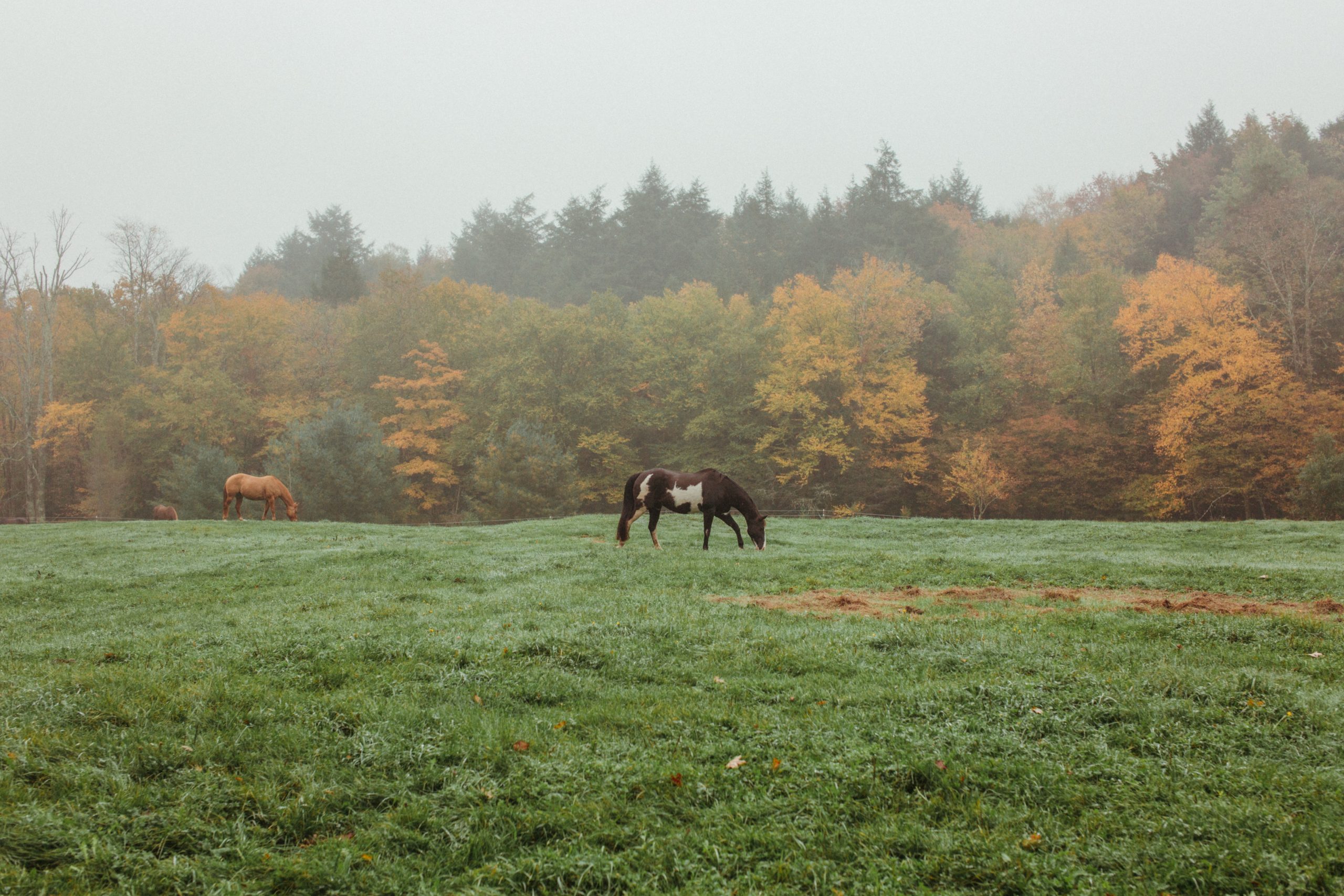
 The arrival of spring means long-awaited opportunities for trail riding, clinics, and horse shows. However, travel, new stabling, and the mixing of horse populations can also provide the perfect conditions for the spread of disease. With a little planning and...
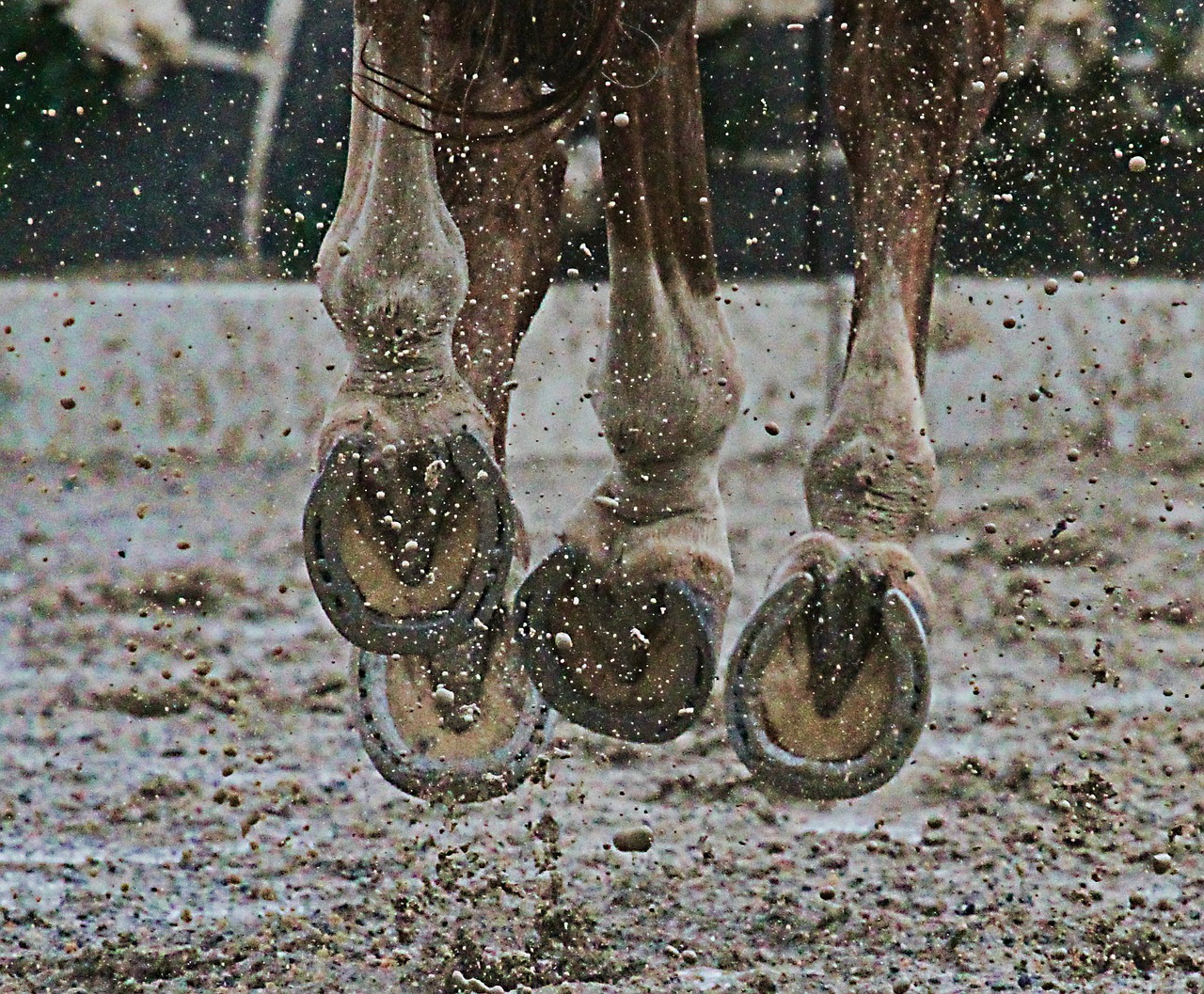 While we are always grateful for precipitation here in Colorado, wet and muddy conditions contribute to the development of two common horse hoof conditions: abscesses and thrush. Abscesses A hoof abscess commonly presents as an acute onset severe lameness without...
 No matter how safe their environment may be, horses will always manage to find trouble. Unfortunately, a not-so-uncommon equine emergency is a sharp object that has penetrated through the bottom of the hoof, also known as a street nail. Any penetrating injury to the...
 Donkeys and mules are readily recognizable by their distinguished large ears. These equids frequently have the reputation of requiring less veterinary attention. Although they are certainly more stoic (and perhaps smarter!) than horses, they still need—and deserve—the...


