Equine Skin Cancers

Skin tumors in horses are not uncommon. While a tissue biopsy is the definitive way to identify the tumor type, location and appearance can offer clues for identification.

Sarcoids

Sarcoids are the most common skin tumor in horses and can be separated into different types depending on appearance.

  • Occult sarcoids are the earliest form of sarcoids and can progress to other forms or remain quiet for years. They vary in size and range from a roundish area of slightly different hair type to a gray hairless circular area. The skin can feel thickened and may resemble a rub from tack or ringworm lesion.
  • Verrucous sarcoids appear gray and scaly or wart-like. They can also become ulcerated and the skin in this area may crack easily.
  • Nodular sarcoids are obvious firm masses which may be attached within the skin or the skin may be separate from the nodules. The axilla (arm pit), eyelid, and inner thigh are common locations for nodular sarcoids.
  • Fibroblastic sarcoids are aggressive and ulcerative in appearance. They can occur anywhere on the horse’s body. The other types of sarcoids can evolve into the fibroblastic type from local trauma and irritation. Fibroblastic sarcoids are further divided into subtypes based on the extent of their attachment to deeper tissue.
  • Mixed sarcoids have characteristics of some or all the types listed above.
  • Malevolent sarcoids are fortunately the rarest form. These sarcoids are highly aggressive and spread extensively throughout the skin, with ulcerative and nodular cords of tumor tissue.

While sarcoids can be locally invasive, they typically do not spread throughout the rest of the body. However, they can be frustrating to treat, become very inflamed, and occur in locations that interfere with tack.

Unlike some of the other types of skin cancers, there does not seem to be a color predisposition to development of sarcoids. The development of sarcoids is the result of an individual horse’s immune system/genetic susceptibility and exposure to the bovine papilloma virus.

The location and type of sarcoid determines the best treatment. Unfortunately, there is not any one treatment approach effective for all sarcoids, and individual sarcoids of the same type may respond differently to the same treatment. Treatment can involve topical chemotherapy or chemotherapy injected into the sarcoid, surgical excision, laser surgical excision, immune therapy, electrochemotherapy, or radiation. Incomplete removal of a sarcoid can lead to recurrence at the same site and a more aggressive sarcoid.
Squamous cell carcinoma

Squamous cell carcinoma (SCC) is the second most common skin cancer in horses. This tumor type develops from skin cells and is seen more commonly on pink skin. Common locations include the eyelids and external genitalia. In addition to horses with pink skin, such as Appaloosas and paints, Belgians and Haflingers may be predisposed to SCC.

Early SCC can appear as a depigmented patch of skin or dry crusting area, progressing to an ulcerated, raised, or cauliflower-like mass. It is best to be proactive about these lesions early, since removal becomes more challenging with increasing size. Always closely monitor areas of pink skin on your horse.

Treatment options for SCC include surgical removal of the mass, local treatment with chemotherapy, CO2 laser treatment, and cryotherapy. Larger and more complex masses, or those involving significant portions of the eye or eyelid might require removal in a hospital setting by a surgeon. At-risk horses benefit from wearing a UV-protective sheet when UV-exposure is high, as well as a UV-protective fly mask. Remember to check your horse’s eyes daily when wearing a fly mask.

Melanomas

Although human melanomas are frequently associated with UV-exposure, melanomas in horses are usually associated with coat color, with up to 80% of gray horses developing melanomas at some point during their lifetime. Melanomas arise from melanocyte cells, which produce the pigment of the skin. Depending on tumor location, melanomas can dramatically impact the horse’s quality of life. At times they may ulcerate and exude a tarry dark substance (melanin).

Common locations of melanomas include the underside of the tail, around the rectum and external genitalia, and around the mouth. Melanomas can also spread to internal organs, such as the liver, spleen, and lungs. Surgical removal of external masses when they are small is recommended, and surgical removal is more effective than medical therapies.

Medical therapies include local injection with chemotherapy, cimetidine (an antihistamine, with mixed results), and a newer option: a melanoma vaccine. A canine melanoma vaccine has already been developed, so similar research is underway for an equine melanoma vaccine. The idea is to create a vaccine unique to that horse to stimulate the immune system to target melanoma cells. While the vaccine does show some promise in reducing tumor size, it unfortunately isn’t a guaranteed cure.

Bottom line: be proactive in monitoring your horse’s skin, and contact your veterinarian if you observe any suspicious skin lesions.

Top of Form

EGUS Series Part 1: What’s the fuss over gastric ulcers?

Be it in the show ring or over dinner with horse friends, the topic of gastric ulcers always seems to elicit sentiments of frustration and misunderstanding.  What causes these pesky ulcers and what can we do as horseman to combat them?

To start, it’s important to understand that before horses were domesticated by humans, they spent their days slowly grazing across the plains and grasslands.  The slow ingestion of fibrous plant products creates an integral part of the horse’s digestive mechanism called a fiber mat, which naturally floats on the top of the stomach contents. 

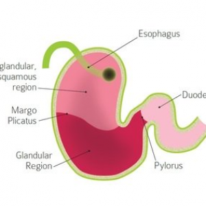
Why is this fiber mat so important?  The horse’s stomach is separated into two regions, glandular and non-glandular.  The glandular region is responsible for the production of gastric acid which is necessary for the digestion of forage.  To avoid self-destruction, the glandular region of the horse’s stomach has protective mechanisms that keep the gastric acid from digesting the very tissue that produces it.  Unfortunately, the non-glandular region does not possess such protection and is susceptible to the destructive effects of the acidic stomach contents.  This is where the fiber mat comes into play.  Horses that graze throughout the course of the day are continuously adding plant fiber to the fiber mat which serves as a protective boundary between the acidic gastric contents and the non-glandular portion of the stomach. 

Let’s jump ahead to our typical domesticated horse which lives in a commercial boarding situation.  These horses are usually fed twice a day, often going more than twelve hours between their evening meal and breakfast.  The detriment here is without the continual ingestion of hay, that fiber mat is quickly digested and sent past the stomach into the rest of the GI tract.  This leaves the non-glandular portion of the stomach exposed and susceptible to the deleterious effects of the acidic stomach juices, as these secretions are produced on a continual basis.  Now, let’s say we decide to ride this horse without his fiber mat and the increased activity level causes more splashing of the stomach contents onto that already sensitive region.  As the acid begins to eat away at the lining of the non-glandular region of the stomach, the horse is thrown into a downward spiraling scenario that, without medical intervention, is quite difficult to recover from. 

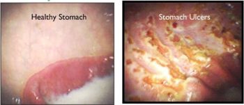
How do you know if your horse has ulcers?  Common clinical signs of a horse with ulcers are aversion to tightening of the girth, sensitivity to grooming (especially behind the elbows), finicky eating habits or changes in behavior both on the ground or under saddle.  The only guaranteed diagnostic method to confirm the presence and severity of gastric ulcers is by passing an endoscope into the horse’s stomach and visualizing the integrity of the gastric mucosal lining.  For many of our clients, this is not a viable diagnostic mechanism, so instead, we assess the horse’s response to medical therapy for ulcers.  If there is a vast improvement in clinical signs over the course of therapy, the likelihood of gastric ulceration is high and we can continue with medical intervention and nutritional management. 

Stay tuned next month for part 2 of our blog discussing the treatment and management of horses with Equine Gastric Ulcer Syndrome (EGUS)!

Should my horse get a strangles vaccine?

Most horse people are familiar with the dreaded “s- word”: strangles. But if there is a strangles vaccine available, why isn’t vaccination essential for all horses, like the rabies vaccine?

What is strangles?

Equine strangles is caused by infection with the bacteria Streptococcus equi var equi, causing mild to severe upper respiratory infection. Complications from infection can occasionally be fatal, but most horses fully recover. Strangles is a high morbidity disease, meaning it is extremely contagious in susceptible populations.  Proper biosecurity measures are crucial to limit the scope of an outbreak.

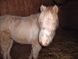
Pony with Strangles shows swelling from abscessed lymph nodes

Signs of strangles include fever, swelling of the lymph nodes (especially in the throat latch area), loss of appetite, cough, and significant mucoid yellow nasal discharge. The bacteria mobilize to the lymph nodes and causes intense immune response, so frequently these lymph nodes abscess open and drain. The term strangles originated from the harsh respiratory noise heard when severe swelling and lymph node abscesses can impair the horse’s ability to breathe.

Most infected horses respond well to supportive care. Supportive measures include anti-inflammatories, hot-packing of abscesses, and rest. More severe cases may benefit from hospitalization and antibiotics. To prevent the spread of strangles, hospitalized cases are managed in the isolation unit.

How does strangles spread?

Strangles is easily spread by horse-to-horse contact and via contaminated surfaces, such as handler clothing and hands, grooming equipment, and water troughs. Most horses will clear the infection within 3-6 weeks, but it is possible for some horses to recover fully and remain shedders of strangles. These horses appear healthy but will shed the bacteria and continue to infect other horses. Upon recovering from strangles, horses will be protected against reinfection for variable periods of time, sometimes even a few years.

Strangles vaccine options

Two types of strangles vaccines are available. One is a “killed” vaccine, meaning it contains dead/inactivated strangles. This vaccine is administered intramuscularly.  Killed vaccines produce a weaker immune response, so it is still possible for a horse vaccinated with this vaccine to develop strangles if exposed. Per the American Association of Equine Practitioners, the killed vaccine should not be expected to prevent disease. It may be effective in lessening the severity of infection. There is also an increased risk of vaccine site reaction with this product.

The second type of vaccine is called a “modified live” vaccine. This vaccine type uses a weakened form of strangles to stimulate a stronger immune response. In order to accomplish this, the vaccine is administered intranasally to target the same tissues as natural strangles infection. However, it is also possible for this modified live vaccine to cause lymph node abscesses, much like natural strangles infection. Horses that have natural immunity to strangles (those who have recovered from infection) have a higher risk of adverse reactions to strangles vaccination.

Should my horse have the strangles vaccine?

The take-home point is that we currently lack a perfect strangles vaccine. If your horse is in a high-risk population, discuss the pros and cons of strangles vaccination with your veterinarian to evaluate if strangles vaccination is worthwhile. It is important to remember that vaccination does not guarantee that your horse will not get strangles. Proper biosecurity is essential to prevent and limit strangles outbreaks.

Equine Cushings Disease

Most horse owners have heard about equine Cushings disease and are familiar with some of the common clinical signs: a long curly haircoat, delayed shedding, topline loss, pot-bellied appearance, and sometimes laminitis. Additional signs include recurrent infections, delayed healing, increased water intake and urination, and lethargy. Especially as our equine companions live longer than ever before, a diagnosis of Cushings disease is very common. But what causes these clinical signs and what can be done for horses with this condition?

What is Equine Cushings/PPID?

Equine Cushings is the most common endocrine disease in our older horse population. Cushings in humans and dogs differs from the equine condition, so equine Cushings is more accurately known as PPID (pituitary pars intermedia dysfunction).

The small pituitary gland is found at the base of the brain near the hypothalamus. It is composed of three different parts, each with unique functions. As indicated by the name, PPID affects the pars intermedia portion of the pituitary gland. The neighboring hypothalamus helps regulate the pituitary’s secretion of hormones. When this regulation is disrupted, the pituitary continues to secrete hormones unchecked. Usually the hypothalamus releases dopamine, a hormone, to signal the pars intermedia to stop producing hormones. In older horses and PPID affected horses, there is less dopamine to inhibit the pars intermedia, so it continues releasing hormones and increases in size. The increased amounts of these hormones, including one called ACTH, affect your horse’s thirst, thermoregulation, and response to stress.

Photo by Moriah Wolfe on Unsplash

How to Diagnose PPID

Diagnosis of PPID is made with blood work to measure the level of the ACTH hormone. Completion of full metabolic panel in addition to ACTH level is important to investigate other hormones that may also be affected, such as insulin and leptin. One important consideration is the time of year when blood is drawn, as ACTH levels of all horses increase during the transitional fall period (approx. mid August through end of November).

PPID Treatment Options

PPID is a manageable, but not curable condition. The mainstay of treatment of PPID involves daily administration of pergolide (prascend). This medication helps reduce ACTH levels and improve clinical signs. Horse owners report improvement in shedding, better maintenance/building of topline, and improved attitude/appetite. Treatment of PPID can also help reduce circulating insulin levels in those horses that experience elevated insulin secondary to PPID, thus reducing the risk of laminitis. For horses with concurrent insulin dysregulation, additional medications and dietary management may be indicated to further mitigate the risk of laminitis. Many herbal remedies claim to aid in treatment of PPID, such as chasteberry, but studies have failed to prove any benefits of supplementation. Treating PPID with pergolide improves both quality and length of life for affected horses.

What’s All the Talk About Insulin? Hyperinsulinemic Associated Laminitis

Thirty years ago, laminitis was usually a career-ending diagnosis, if not a life-ending one.  Aside from mechanical support and pain management, little was known about the contributing factors associated with laminitis and the multitude of clinical factors that affect the severity, outcome, and likelihood of recurrence.  Today, we know a great deal more about metabolic laminitis and specifically the role that insulin plays in mediating this disease process. 

Current data shows that horses with an elevated baseline insulin value are at a higher risk of developing hyperinsulinemic associated laminitis (HAL).  The ability to establish a direct relationship between elevated serum insulin levels and the onset of laminitis has enabled veterinary practitioners to make great strides in combating this disease process. 

Hyperinsulinemic Associated Laminitis Diagnosis and Treatment

When presented with a patient suffering from HAL, the first step is to determine just how high the insulin values are.  Seasoned practitioners can usually estimate the severity of hyperinsulinemia by assessing the extent of regional adiposity (fat deposition in the horse’s body).  The most common site of regional adipose tissue deposition are the crest of the neck, on either side of the withers, along either side of the horse’s topline, and on either side of the tail head.  The greater the accumulation of fat in these areas, the higher the baseline insulin value in that patient.  Bloodwork, in the form of a metabolic panel, is then used to quantify those observations to establish an appropriate therapeutic regimen and track progress throughout the treatment period. 

If the patient is actively suffering from HAL, all therapeutic methods are implemented in an attempt to slow down the damage associated with the laminitic process.  In addition to dietary management and the eradication of starch from the horse’s feed, medical intervention with metformin has proven to be a very successful strategy in our practice. 

Metformin increases tissue sensitivity to insulin in the patient. Insulin is a signaling molecule which instructs cells to recover glucose (starch/sugar) from the GI tract to use to power cellular processes.  In horses with hyperinsulinemia, the tissues of the body aren’t responding to the insulin currently being produced, so, the body produces more insulin, leading to a hyperinsulinemic state. 

The physiologic process by which elevated insulin values lead to laminitis are still unknown.  However, current research shows that insulin is capable of binding to receptors in lamellar epithelial cells which stimulates excessive growth of the horn tubules, leading to the traditional elongated hoof structure of chronically laminitic feet.  Metformin helps to increase tissue sensitivity to insulin which in turn down regulates the body’s natural production of insulin. 

Hyperinsulinemic Associated Laminitis Treatment Outcomes

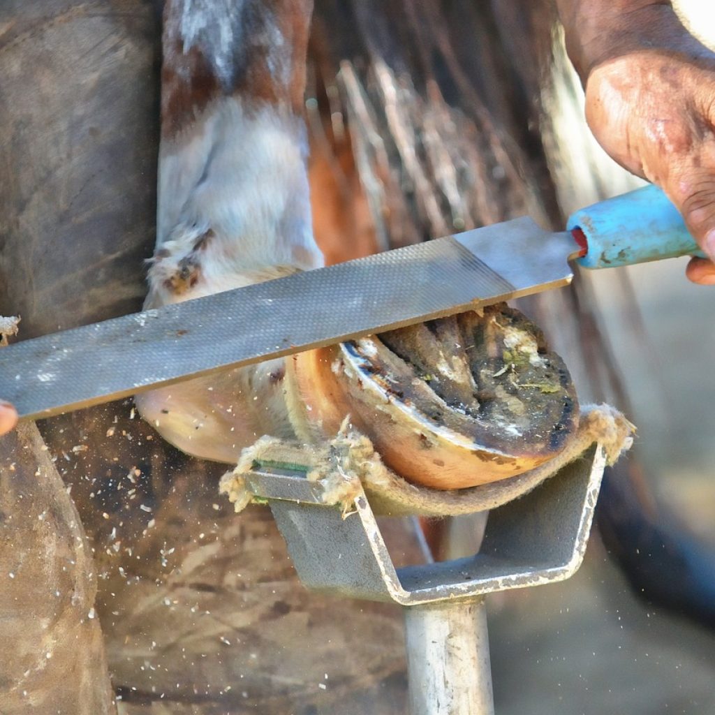
The success of treatment of horses with insulin dysregulation is highly dependent upon the severity of the HAL, the chronicity of the disease process, and the condition of the hoof capsule.  Optimal outcomes are associated with high compliance on the part of the horse owner when it comes to implementing therapeutic regimens and dietary management, as well as the employment of a farrier who is willing to work with your veterinarian when making strategic decisions around trimming and shoeing your horse. 

These patients require several series of radiographs over the course of their case in order to ensure optimal trimming is being performed in addition to tracking the sole depth of the patient.  Most cases, when identified in their chronic stages, usually require many months to restore physiologic function of the hoof capsule and achieve an acceptable level of comfort on the part of the patient.  Depending upon the integrity of the hoof capsule, metabolic stability, and comfort of the patient, these patients can sometimes return to their previous level of work.  While not all cases have the perfect outcome, with the knowledge and medical advancements the veterinary profession has seen over the past few decades, it is absolutely worth trying to combat hyperinsulinemia associated laminitis. 

Horse Show/Travel Biosecurity

The arrival of spring means long-awaited opportunities for trail riding, clinics, and horse shows. However, travel, new stabling, and the mixing of horse populations can also provide the perfect conditions for the spread of disease. With a little planning and biosecurity awareness, you can minimize potential risks to your horse.

What is Biosecurity?

Biosecurity refers to the preventative measures taken to minimize spread of disease. The most commonly encountered infectious diseases include influenza (flu), rhinopneumonitis (equine herpes virus), and strangles. While many horses are vaccinated against these diseases, vaccination does not guarantee 100% protection and does not negate the need for biosecurity.

How do I take effective biosecurity measures?

Some biosecurity measures are common sense – such as not allowing your horse to have nose-to-nose contact with other horses. Other measures are more complex.

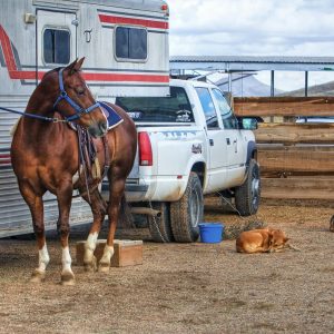
First, consider your destination. Horse show venues and campgrounds may have specific requirements for incoming horses, such as a current Coggins test, health certificate, and proof of up-to-date vaccination. Remember to keep your veterinarian apprised of your summer travel plans so that you have all required documentation prior to your departure. While it can be frustrating to manage differing requirements, remember they are put in place for the health and safety of the horse.

When arriving at a campground or show stable, examine the housing for your horse prior to unloading. Look for overall cleanliness – an area free of manure and old bedding. It’s worth asking the venue how facilities are cleaned between horses prior to your arrival. Dirt floors and other porous surfaces are much harder to disinfect since organic debris can inactivate cleaners such as bleach.

Simple and easy biosecurity measures:

  • Prevent all direct contact between your horse and others (no nose-to-nose!)
  • Avoid sharing equipment with others, including; tack, grooming supplies, wheelbarrow, pitchfork, etc.
  • Avoid communal water sources/sharing hoses between horses. Dropping the end of the hose into multiple water buckets can transmit disease.
  • Wash your hands frequently and after touching any other horses.
  • Pack your horse’s thermometer. If your horse appears “off” a fever may be present before he is contagious to others.
  • Don’t forget to clean and disinfect your trailer regularly too!

Wishing you a safe, happy, and healthy riding season!

Emerging Infectious Diseases: What you need to know

Ten years ago, it was quite uncommon to order diagnostic testing for Equine Protozoal Myeloencephalitis (EPM) in horses demonstrating signs of neurologic disease in Colorado.  Additionally, it was rare to evaluate equine patients for Lyme Disease in Western states.  That all appears to be changing. 

Lyme Disease

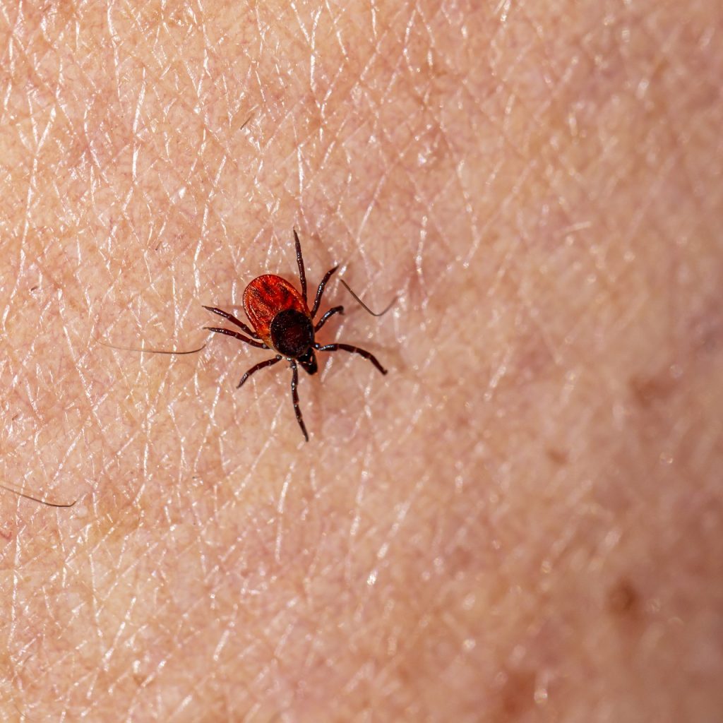
Lyme Disease is a tick transmitted infection caused by the bacteria Borellia burgdorferi, and to a lesser extent, Borellia mayonii.  Historically, only the tick species Ixodes scapularus and Ixodes pacificus(black legged ticks)were known to carry these bacteria, and neither of these species were found in Colorado.  While there have only been seven cases of Lyme Disease reported to the CDC in Colorado since 2019, there is a high probability that a lack of biosurveillance and underreporting have resulted in an underrepresentation of Lyme Disease in the state.  According to the CDC, all seven cases of Lyme Disease in 2019 originated from outside of Colorado.

Black Legged (Deer) Tick

The black legged tick resides mostly along both coasts of the United States, as well as all Midwestern states from as far north as Minnesota and as far south as Texas.  Many of these ticks have been found in Utah as well, Colorado’s most immediate western neighbor.  As our climate begins to change so too will the tick populations within the western states.  The most important thing horse owners can do to protect their horses from tick borne disease is to perform thorough examinations of your horse’s legs and underbelly, neck, armpits, and chest for any evidence of ticks.  Initially they may feel like a small growth or skin tag, but upon further examination, it will become apparent that it’s actually an external parasite.  Care should be exercised when removing these creatures from your horse’s skin to ensure that the head is grasped tight enough to facilitate removal along with the body but not too tight to cause premature squishing of the tick prior to detachment.  Tick bites usually result in a local inflammatory response so evidence remains even after natural detachment in the form of a small scab. 

Testing for Lyme Disease is via a simple blood test which is then sent to Cornell University.  The current test in use is called a Lyme MultiPlex Assay and enables us to differentiate between acute and chronic infections.  This information in turn is used to pursue the optimal treatment option for each individual case.  Horses can never be cured of Lyme disease, but through prolonged treatment regimens, clinical symptoms can often be put into remission.  Patients with previous exposure to Lyme disease are always susceptible to relapse.  Symptoms of Lyme disease include lethargy, a history of poor performance, ventral limb edema, shifting leg lameness and a multitude of other nonspecific clinical signs.

Equine Protozoal Myeloencephalitis

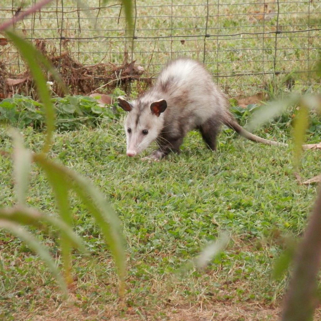
Equine Protozoal Myeloencephalitis (EPM) is another disease that is associated with a varying array of clinical signs.  Mild symptoms can be as vague as a history of poor performance.  More severe signs are associated with neurologic deficits and can even progress to recumbency.  EPM is found in the feces of opossums infected with the protozoa parasites Sarcocystis neurona and Neospora hughesi.  When horses ingest the feces of infected opossums, there exists a significant risk of the establishment of infection with the protozoa in the horse’s central nervous system.  Currently, the opossum population in Colorado does not carry the protozoa responsible for EPM, but, horses that travel outside of Colorado into EPM endemic areas are at an increased risk of exposure and development of EPM. 

Like Lyme Disease, horses infected with EPM will always have a risk of relapse of clinical symptoms, even after extensive therapy.  There is no permanent cure.  Diagnosis is achieved through blood testing as well as the acquisition of a sample of cerebral spinal fluid (CSF).  CSF is more specific than blood in confirming a diagnosis of EPM but the risks associated with the collection of a CSF sample are far greater.  Treatment of EPM does not offer a cure for the disease but instead aims to decrease the severity of clinical signs.  Usually, we anticipate an improvement of one grade of ataxia on Mayhew’s grading scale for ataxia after completion of a therapeutic regimen.  As such, if a horse has a starting ataxia grade of 1/5, the prognosis for return to work after treatment is optimistic.  If a horse is starting at an ataxia grade of 3 or 4/5 prior to treatment, the prognosis for a full return to work is much more grave. 

While we are seeing an increase in both Lyme disease and EPM in our patients, the risk of contracting either disease if your horse never leaves Colorado is still quite low, but not impossible.  We as equine practitioners will continue to think outside the box and ensure that all possible explanations for your horse’s clinical signs have been pursued and ruled in or out. 

Is your horse current on his rabies vaccination?

Rabies is caused by a virus that infects mammals and is spread through the saliva/bites by infected animals. The virus exists in several wild animal reservoirs, most commonly bats, skunks, raccoons, and foxes. After the virus is transmitted via bite, it replicates in the muscle at the site of the bite, and then travels to the central nervous system (brain and spinal cord). The virus continues to replicate in the central nervous system and then spreads through the rest of the body, including the salivary glands, which then allows transmission to other susceptible animals.

Is rabies a problem in Colorado?

In 2019, there were 173 lab-confirmed cases of animal rabies in Colorado, including canine, feline, and camelid cases. So far in 2020, Colorado has seen 71 lab-confirmed cases of rabies, including in a dog, goat, sheep, and bull. It is important to remember that because rabies is endemic in bat, skunk, and raccoon populations, these numbers don’t accurately represent the true prevalence of this disease.

What does rabies look like in horses?

While mention of rabies conjures images of a frenzied animal frothing at the mouth, clinical signs in the horse can be extremely variable, so many people may be potentially exposed before a diagnosis is made. Non-specific signs of rabies in horses can range from colic and lameness to fever and depression. There is no available testing to diagnose rabies in the live horse. When examining an un-vaccinated horse exhibiting bizarre behavior or neurologic signs, your veterinarian must consider rabies as a potential diagnosis. Signs in an infected horse progress rapidly, usually leading to death within 5-7 days. Rabies is always fatal in an unvaccinated horse.

The rabies vaccine is a core vaccine for horses, according to the American Association of Equine Practitioners (AAEP). A core vaccine is one that is essential for every horse, regardless of the horse’s lifestyle. Rabies is a FATAL disease but it is preventable through annual vaccination by your veterinarian. Because human infection is also fatal, rabies vaccination in domestic animals is a matter of public health. Any person who may have potential exposure to rabies should immediately contact their health provider.

Protect your horse- and yourself! Be sure to keep your horse’s rabies vaccination current, and don’t forget to vaccinate your barn cats and dogs!