Caring for Your Senior Horse

At what age is a horse considered geriatric or senior? Does owning a senior horse mean that he must eat a feed labeled as a “senior” feed? As always, the answer is, it depends. Our horses are living longer lives than ever, thanks to an evolving understanding of the needs of geriatric horses and our ability to provide high quality care. There is no set age cutoff as to when a horse is considered geriatric, but most experts agree this can be around age 20. However, age is just a number, and many horses are still active and competing into their twenties, while others are happily retired. The main issues we see in our older horse population include dental disease, altered dietary needs, metabolic disease/PPID (Cushings), and arthritis.
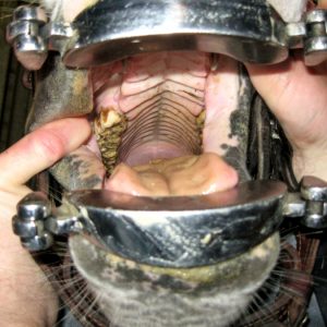
 

Photo Courtesy PugnoM on Flickr

Dental Changes

The ideal time to begin focusing on your horse’s regular preventative veterinary care is when he is young. Horses receiving regular preventative care, especially veterinary dentistry, preserve their teeth longer and can age more gracefully. Waiting until signs of dental disease or difficulty chewing are observed often indicates advanced disease and more limited treatment options. Most horses benefit from yearly dentistry, which includes a thorough sedated oral exam with a dental speculum, light source, and mirror. A thorough oral exam is vital to accurately assess, document, and treat any issues.

Horses suffering from abnormal dental wear, cracked or missing teeth, or severe periodontal disease may require more frequent care. Even though horse teeth are described as “long rooted,” they do eventually wear out, so proper care ensures the longest possible life of the tooth. Common dental issues in older horses include fracture of cheek teeth, complete expiration or wearing out of cheek teeth, loose/painful teeth, and periodontal disease. The incisors can also be affected by similar conditions.

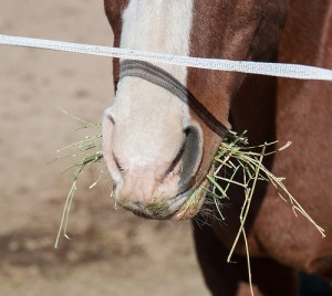
Staying on top of your senior horse’s dental care helps ensure that he gets what is needed from his diet. Sometimes severe dental disease and impaired chewing ability necessitates eliminating hay from the diet entirely. Complete feeds are formulated specifically for horses that can no longer chew hay effectively. These diets have greatly improved the quality and length of life for senior horses with compromised teeth.

Nutritional Changes

Nutrition recommendations for the geriatric horse are formulated around maintaining an ideal body condition. In the geriatric horse, this can be complicated by difficulty chewing, poor teeth, metabolic disease, and decreased ability to digest fiber and protein. For older horses in good weight and with adequate dentition, little dietary change may be required. For older horses who have difficulty maintaining weight and/or compromised teeth, complete feeds as mentioned above can help. These feeds are high in easily digestible fat, fiber, and protein, and are designed as easy-to-chew. They are formulated to replace hay entirely for those horses that can no longer adequately chew hay.
Adding water to soften the feed can reduce the risk of choke and ensure additional water intake. Feeding recommendations are often listed as pounds of feed per day depending on whether the horse is also eating hay, so it is important to weigh out your horse’s portion so that any necessary adjustments can be made more accurately.

Metabolic Changes

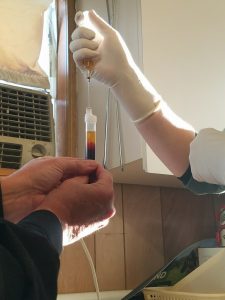
A curly haircoat and muscle loss over the back aren’t just signs of aging- They can indicate a common underlying condition: equine pars 
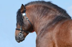
pituitary intermedia dysfunction, more commonly known as PPID or Cushings disease. This is a treatable (but not cureable) condition that is very common in geriatric horses, resulting in hormonal imbalances and symptoms such as delayed shedding, curly hair coat, muscle loss over topline, increased drinking and urinating, personality changes, and increased susceptibility to infections and laminitis. Diagnosis is made by a blood test as well as by history and clinical signs. Management involves daily administration of pergolide (Prascend) tablets, which help restore normal hormone levels and alleviate the clinical signs of the disease.

Equine Metabolic Syndrome (EMS) is a separate metabolic condition, but it sometimes occurs in conjunction with PPID. EMS horses typically show signs of being an “easy keeper”- cresty neck, regional fat deposits behind the shoulders and at the tail head. Horses with EMS are more susceptible to laminitis and usually have a higher baseline insulin level than normal. EMS horses are managed with strict attention to diet (minimizing sugar and starch) and exercise to help them maintain a lean body weight.

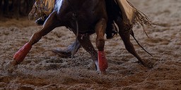
Joint and Muscle Changes

Just like us, aging horses suffer from daily aches and pains. An examination with your vet can help identify major and minor issues and

Photo Courtesy nikki_tate on Flickr

determine what treatment plan works best for you and your horse. Many options are available to keep our old horses comfortable.
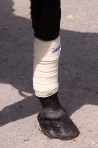
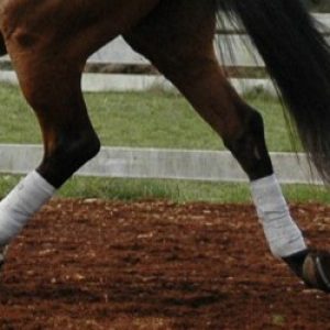
Nutraceuticals (supplements) are very popular, although many of these products lack proof of efficacy and ingredients. More researched options include medications such as adequan (administered intramuscularly) and Legend (administered intravenously). These medications help provide the components necessary to keep joints healthy.

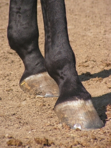
Joint injections target individual joints to reduce pain and inflammation and improve mobility. Non-steroidal anti-inflammatories such as Equioxx are also helpful, along with acupuncture and chiropractic care. Stable management is equally important- maximum turn out time allows horses to move at will, and monitoring herd dynamics ensures that older horses are not prevented from accessing food and water. In addition to these considerations, regular veterinary and farrier care will help your senior horse enjoy his golden years with you.

As always, if you have any questions about caring for your senior horse, please contact your veterinarian who can offer a personalized plan to help keep your geriatric horse happy, healthy, and comfortable well into their senior years.

Joint Injections: The what, when and why of intraarticular medications

Photo Courtesy nikki_tate on Flickr

Arthritis is one of the most common joint diseases found in the equine industry.  It is the leading factor of decreased performance and chronic lameness in our equine partners and is the source of much frustration and consternation.  The insidious nature of the process means that we as horseman don’t always catch it in the early stages, and instead, are left dealing with the disease in more moderate to advanced situations.  In a previous blog we’ve focused on how to prolong the onset of arthritis by using systemic nutraceutical or injectable products like Cosequin and Adequan (you can find that post here).  This article will focus on the options available to us when these initial attempts against arthritis are no longer efficacious.

What happens during a joint injection?

Once our systemic therapies like Adequan and Legend have no longer proven to be effective in reducing joint pain associated with arthritis, we turn to localized treatment directly within the damaged joint.  Prior to injecting any joints, your veterinarian will perform a thorough lameness exam to isolate the joint(s) from which the lameness is originating.  From there, we as veterinarians prefer to obtain radiographs of the joint in question.  This provides us both a baseline for future assessments as well as a better opportunity to understand any changes to the diseased joint that may alter our approach to needle placement.  The information obtained from the radiograph enables us to give our clients a more accurate prognosis for both post-injection soundness and duration of efficacy of the injection.

Preventing joint infection

Once you and your veterinarian have agreed to inject a joint(s), your veterinarian will then display an odd set of behaviors in which they tirelessly seek out the ideal location to perform an injection.  The biggest risk associated with a joint injection is infection, so we tend to be pretty picky about when and how we inject.  We avoid all windy situations, including drafts, as it seems just as you place the needle into the joint, a large gust of wind comes along and stirs up dust and debris which could potentially contaminate the joint.  We also avoid injecting horses with wet mud/manure directly over and surrounding the site of injection.  Clean, dry legs are necessary. We avoid areas of high traffic, as all it takes is an unknowing rider or the sneeze of a horse to contaminate our sterile site.

The ideal environment for a joint injection is one that has clean, dry footing (rubber mats or concrete work best), that is enclosed to restrict

Image Courtesy USMC via Flickr

or prevent airflow.  Fewer birds in the overhead peanut gallery is also preferred!  If these requirements cannot be found, don’t be surprised if your vet chooses to reschedule for a calmer/dryer day or you’re asked to relocate to a more ideal location.  At the end of the day, a joint infection can be life altering so we strive to avoid those at all costs!

The injection process itself is also sterile.  We will relocate all sources of potential contamination (tails, lead ropes, etc.) and will usually clip the injection site.  All joint injections are preceded with a 12-minute sterile scrub, and all products used in the injection process are also sterile, including your vet’s gloves!  Most practitioners will also use a small amount of antibiotic within the injection itself to further reduce the risk of infection.

What exactly is injected into the joint? 

As mentioned before, there is a small amount of antibiotic that is included in every injection, but beyond that, there is significant variability.  Larger volume joints (think coffin joints, knees, stifles, fetlocks) usually receive a high molecular weight hyaluronic acid (HA) and possibly steroids.  The steroids work to decrease the pre-existing inflammation within the joint and the HA helps bring healthy components into the area to improve the overall joint environment.  (There is also a mild anti-inflammatory response associated with HA).  Lower volume joints (pastern joints and the lower hocks) usually just receive steroids and antibiotic, forgoing the HA due to the small volume of joint space available to accommodate the injection.  There are several types of steroids available to use and which steroid we use is dependent on a multitude of factors.  These include high vs low motion joints, age of horse, severity of the intraarticular disease process, and metabolic status of the horse.  Steroids are contraindicated in horses with metabolic disorders which then has us reaching for an alternative – autogenous intraarticular therapies also termed “regenerative medicine.”

Regenerative Medicine Therapies

For those horses with metabolic issues or who no longer respond well to intraarticular steroids, we now have a variety of options for use in their place.  IRAP (interleukin-1 receptor antagonist protein), PRP (platelet rich plasma) and Prostride are all newer, autogenous technologies available to the veterinary industry.  While PRP and ProStride have applications in horses with arthritis, IRAP is far and away the most commonly used.

IRAP is processed directly from your horse’s blood and it works to counteract the interleukin-1 (IL-1) inflammatory protein.  The blood is collected into a special tube that is then incubated for several hours.  During the incubation process, the IRAP molecule is amplified within the sample.  After incubation, the sample is spun in a centrifuge which then separates the red blood cells from the IRAP rich plasma.  The plasma is then divided up into small aliquots to be used during the joint injection process.  Unlike steroids, which usually only require one injection to have a beneficial response, IRAP requires 2-3 injections, separated by 2 weeks in order to deliver similar results.  The IRAP injections usually last an average of 6-8 months, but the duration of efficacy is dependent upon a multitude of factors and is different for every horse.

While arthritis can be a difficult condition to overcome, rest assured that we do have a large variety of therapeutic options to pursue.  Please don’t hesitate to reach out with additional questions or concerns the next time you see one of us at your barn.  We’re always happy to help you and your horse!


Lacerations- When do you need to call the veterinarian?


By Dr. Shane Baird

HorseWoundAs horse injuries go, cuts, scrapes, and bruises of all shapes and sizes are common – the challenge is learning when veterinary intervention is required.

Is There Joint Involvement?

One of the most critical factors for laceration severity is the location of the laceration.  A solid “rule” to keep in mind is that if the wound is over an area that “bends” and appears to be fully through the skin, it justifies at minimum a call, and likely a visit from a veterinarian.  Cuts that seem to be no big deal, but are located directly over a joint, can be disastrous for the horse, while something that looks horrible at first glance but only involves skin and muscle can heal without causing any problems.

The lower limbs of horses don’t have a lot of tissue between the skin and the joints or bones. This “lack of padding” makes it easy for a cut that looks simple to penetrate into the joint.  Joints open to the outside world are open to infections and if infected, can become debilitating for a horse due to the arthritis associated with the process.

Severe Wounds Without Joint Involvement

Severely traumatic wounds, where tissue (skin, muscle) have been damaged or are just missing, can greatly benefit from prompt veterinary attention.  Proper removal of tissue and thorough cleaning of these wounds will speed the healing process significantly.  Though some of the skin may be missing from a wound like this, with some creative suturing, what remains can be used as a natural bandage, even if only temporarily.  At times, if healing is taking a longer than expected, there is justification for skin grafting techniques, even in an ‘in the field’ situation.

Head Woundshorse-943771_1920

Though the head can heal quite well on its own, lacerations to the eyelid region and around the eyes often warrant closer examination.  If your horse has an injury like this and they have no desire for you to even look, much less touch and clean the wound (which a fairly common response!), please call your local veterinarian so that things can be tended to (likely with sedation).  Trying to fight with the horse can quickly lead to other issues or injuries.  With sedation these wounds can be treated without further injury to the horse or any humans.

Tips on Pre-Veterinary Visit Wound Care

bandage on the leg of horse

If you do decide that a veterinarian should see the laceration, here are some basic rules to follow:

  • Do NOT wash the wound with the hose. This can force dirt and debris deeper into the wound.  Using a paper towel or gauze with dilute betadine, clean the wound of obvious dirt and debris.  If the wound has potential to get dirty again before the veterinarian arrives, place a light bandage over the wound.
  • If there is a chance that the wound will need suturing or stapling, DO NOT use anything “blue or purple in color.” While efficient at stopping bleeding, these dusts and powders can be very damaging to healthy tissue and create a larger mess than before.
  • If your vet is not going to see the wound, and it doesn’t need a full bandage, a little bit of ointment (triple antibiotic is good) will help to keep the wound clean and moist.
  • If bandaging, make sure there is “padding” between the skin and any “vetwrap” like product.

As always, if there is any question, give your veterinarian a call. After a conversation you can both make a decision of whether your horses’ laceration should be seen or not.


Equine Metabolic Syndrome and all it Entails

By Dr. Allison Hartman


the-hoof-473095_1280Laminitis has been a bane of horseman and farriers since the horse was first domesticated.  As our medical knowledge and diagnostic abilities have evolved, so has our understanding of the contributing factors of this disease process, including Equine Metabolic Syndrome.  Equine Metabolic Syndrome is a term that’s been thrown around the equine community for the past few decades, but what does it mean?


What is EMS?


Equine metabolic syndrome (EMS) is a condition most commonly characterized by an inability to properly metabolize carbohydrates. The disease has been known by many names, including hypothyroidism, peripheral Cushing disease and pre-laminitic syndrome. Today, we know that Equine Metabolic Syndrome is characterized in horses, ponies and donkeys by obesity, regional deposition of fat, and systemic insulin resistance.Shetland_pony_dalmatian2


Insulin is an important hormone that allows the cells to obtain glucose from food.  Without insulin, or a proper response to insulin, cells cannot utilize glucose, regardless of how much food the horse is fed.  This starvation process at the cellular level pushes the horse into a stressed state (characterized by elevated cortisol levels) where the body is encouraged to hold on to as much fat as possible, making weight loss next to impossible in these horses.  In addition, glucose deprivation in the laminae of the horse’s feet can lead to laminitis.
How is it Diagnosed?


When we suspect a horse may have EMS, we will recommend bloodwork to confirm our suspicions as well as guide us in the proper treatment of your horse.  Horses with EMS may also have concurrent Cushing disease, which has been shown to be a contributing factor to the initial development of EMS.  It is important for us to understand the underlying metabolic factors that are contributing to your horse’s case in order to provide the most efficacious recommendations both medically and diet.


The key values we assess when running blood work for EMS include:

  • ACTH: When elevated, this value can be indicative of Equine Cushing’s Disease.
  • Insulin: Elevated levels of insulin indicate insulin resistance.
  • Leptin: This is a secondary value, used to ascertain the validity of elevated Insulin. When leptin is elevated in addition to elevated insulin, EMS and IR (insulin resistance) are valid diagnoses.
  • Glucose: When both ACTH and insulin are elevated, glucose can help differentiate the primary disease process.


What Can You Do About It?


Horses that are merely overweight but not actively laminitic should be put on a low starch diet, comprised of hay with less than 12% non-structural carbohydrates fed at 1-1.5% body weight.  (in this example, a 1000lb horse should be fed 10-15lbs of hay per day).  Their forage diet can be adequately balanced using either a ration balancer or a specifically formulated low starch feed.  These horses also benefit from routine exercise as an additional aid for weight loss.


In those patients that are actively laminitic, exercise is not recommended but the same dietary recommendations apply.  In addition, if your horse is diagnosed as insulin resistant (IR), metformin is a beneficial medication used to increase tissue sensitivity to insulin.  Horses with IR are comparable to humans with type 2 diabetes, in that they produce adequate amounts of insulin, but their body simply fails to respond appropriately.  Metformin is a medication commonly used in humans with IR and its efficacy in equine IR cases has been confirmed both in laboratory and field studies.


pony-2472424_1920Another option to stimulate weight loss in EMS horses is levothyroxine, commonly sold in the equine community as Thyro-L.  Thyro-L functions by increasing the rate of the horse’s metabolism to further stimulate weight loss.  It is important to understand that Thyro-L has no direct impact on the laminae of an actively laminitic horse and instead works indirectly over a longer period of time.


EMS is a frustrating disease for practitioners, clients and patients alike.  The more we understand of the disease process the better our chances at restoring metabolic equilibrium and preventing further deterioration of the laminae.  If you suspect your horse may have EMS, talk to your vet about appropriate diagnostic measures.

The Pre- Purchase Exam- Should You have Radiographs or Ultrasounds Taken?

cowgirl-419084_1920By Dr. Shane Baird

After weeks of searching, you have finally found the right horse.  He’s everything you’ve ever wanted.    What now?

A veterinary pre-purchase exam should be the next thing on your mind!  At a minimum, an active and passive exam by an equine veterinarian should be performed prior to purchase, but what about additional diagnostic imaging?  Regardless of whether or not you’re buying your next equine athlete or your trail partner for the next twenty years, radiographs and ultrasound imaging may be in your best interest to protect your investment.

The Right Horse is the Right Horse For You

In all the pre-purchase exams we’ve performed, we have yet to find a “perfect” horse, and a horse that may be right for your goals, might not be the right horse for someone else’s.

It is important to remember that it is not the responsibility of the veterinarian to “pass” or “fail” a horse.  What you should really be asking is, can you and your horse live with the faults identified during the exam and the limitations that arise because of those shortcomings? We are there to provide you, the buyer, with information pertinent to the health and soundness of this horse so you may decide how the horse may fit in your life and barn.

In some situations, it’s easy to determine whether or not the horse in question will stand up to his intended purpose.  In other situations, we must rely on additional diagnostic means to answer these questions, thus enabling you to make a more informed decision.

When Should You Get Digital Images?

lawn-2599037_1920Let’s say that your veterinarian has completed the basic pre-purchase exam and that a few issues have been discovered that require additional consideration.  You may decide that the findings are small enough that they don’t impede your future plans with this horse, in which case the exam may not need to go any further. However, you might decide that the findings could deter you from purchasing the horse.  This is when we would recommend further diagnostics in the way of nerve blocks, in addition to radiographs and possibly ultrasound.

Ultrasound technology is far more portable and convenient to use stall side than it was even just a few years ago.  If there is any palpable soft tissue issue, but no lameness is detected on exam, then a scan is completely justifiable, especially if the horse is responsive to palpation of the area.  Ultrasound can help us to see issues of the past and try to understand if they could become an issue in the future.

Similarly, radiology (x-ray images), are often the quickest way for us to understand a mild lameness that is identified in the dynamic portion of the exam.  Technology these days enables us to develop radiographs within seconds as they appear on a computer screen.  We may determine that there is a mild amount of arthritis in a specific joint or that this horse has significant degenerative changes that may impact his ability to perform as desired, or as long as desired.  Radiographs, and digital/stall side images are the best way to differentiate between the two.

While forecasting the future is always the most difficult part of the pre-purchase exam, advanced diagnostic imaging can make that picture just a little clearer! Awoman-with-horse-2631173_1920 complete exam, and imaging to help understand any issues uncovered, is our best shot as veterinarians to help you, the buyer, and make sure this new horse is truly the “right one for you!”