Should my horse get a strangles vaccine?

Most horse people are familiar with the dreaded “s- word”: strangles. But if there is a strangles vaccine available, why isn’t vaccination essential for all horses, like the rabies vaccine?

What is strangles?

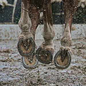
Equine strangles is caused by infection with the bacteria Streptococcus equi var equi, causing mild to severe upper respiratory infection. Complications from infection can occasionally be fatal, but most horses fully recover. Strangles is a high morbidity disease, meaning it is extremely contagious in susceptible populations.  Proper biosecurity measures are crucial to limit the scope of an outbreak.

Pony with Strangles shows swelling from abscessed lymph nodes

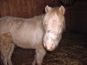
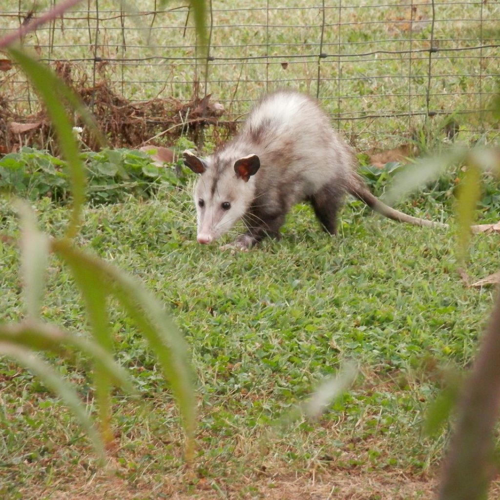
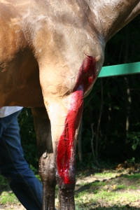
Signs of strangles include fever, swelling of the lymph nodes (especially in the throat latch area), loss of appetite, cough, and significant mucoid yellow nasal discharge. The bacteria mobilize to the lymph nodes and causes intense immune response, so frequently these lymph nodes abscess open and drain. The term strangles originated from the harsh respiratory noise heard when severe swelling and lymph node abscesses can impair the horse’s ability to breathe.

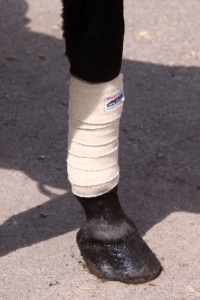
Most infected horses respond well to supportive care. Supportive measures include anti-inflammatories, hot-packing of abscesses, and rest. More severe cases may benefit from hospitalization and antibiotics. To prevent the spread of strangles, hospitalized cases are managed in the isolation unit.

How does strangles spread?

Strangles is easily spread by horse-to-horse contact and via contaminated surfaces, such as handler clothing and hands, grooming equipment, and water troughs. Most horses will clear the infection within 3-6 weeks, but it is possible for some horses to recover fully and remain shedders of strangles. These horses appear healthy but will shed the bacteria and continue to infect other horses. Upon recovering from strangles, horses will be protected against reinfection for variable periods of time, sometimes even a few years.

Strangles vaccine options

Two types of strangles vaccines are available. One is a “killed” vaccine, meaning it contains dead/inactivated strangles. This vaccine is administered intramuscularly.  Killed vaccines produce a weaker immune response, so it is still possible for a horse vaccinated with this vaccine to develop strangles if exposed. Per the American Association of Equine Practitioners, the killed vaccine should not be expected to prevent disease. It may be effective in lessening the severity of infection. There is also an increased risk of vaccine site reaction with this product.

The second type of vaccine is called a “modified live” vaccine. This vaccine type uses a weakened form of strangles to stimulate a stronger immune response. In order to accomplish this, the vaccine is administered intranasally to target the same tissues as natural strangles infection. However, it is also possible for this modified live vaccine to cause lymph node abscesses, much like natural strangles infection. Horses that have natural immunity to strangles (those who have recovered from infection) have a higher risk of adverse reactions to strangles vaccination.

Should my horse have the strangles vaccine?

The take-home point is that we currently lack a perfect strangles vaccine. If your horse is in a high-risk population, discuss the pros and cons of strangles vaccination with your veterinarian to evaluate if strangles vaccination is worthwhile. It is important to remember that vaccination does not guarantee that your horse will not get strangles. Proper biosecurity is essential to prevent and limit strangles outbreaks.

Summer Skin Conditions

What a year here in Colorado!  From fetlock deep mud in the spring to humidity, summer storms and a fly plague of biblical proportion…. No wonder our horses are struggling to keep their skin intact and healthy this year!

When mammalian skin spends a prolonged period subjected to constant moisture, its natural, inherent defense mechanisms become weakened, making them susceptible to any number of dermatological conditions.  For horses this year, the most common skin issue we encountered was pastern dermatitis, commonly known as “scratches”.  This condition presents as an accumulation of scabs in the horse’s pastern region.  It can be quite painful to palpation and even present as a source of lameness.  Once the condition has been identified, the next step is softening the scabs enough so that they can be removed with as little discomfort to the horse as possible. 

My tried and true remedy for scratches is a 50:50 combination of Extra Creamy Desitin (zinc oxide) and nitrofurazone.  (With the mention of nitrofurazone, it is paramount to note that this is the ONLY time I ever recommend using nitrofurazone.  Nitrofurazone has been shown to slow wound healing and tissue regeneration, so I never promote its use on wounds or any other skin lesions).  Once you’ve mixed up a hefty amount of this magical combination product, you’ll want to slather the scabs with a thick coat of this goop and let sit for at least twelve hours.  The zinc oxide is an excellent skin protectant and the nitrofurazone helps to rehydrate the scabs and hamper microbial growth. 

Once the scabs have been softened, you can begin removing as many of them as possible.  Keep in mind that the skin underneath is quite sore and painful so work slowly and gently to keep your horse compliant over the next few days of scab removal.  Once you’ve removed as many scabs as possible that day, gently scrub the area with betadine scrub, rinse, dry and reapply the magical goop.  You’ll repeat this process every 24 hours until all the scabs have been removed.  At that point, it is safe to cease the betadine scrubs and just rinse, dry and reapply a light layer of the magic goop for another 5-7 days until the skin is soft and comfortable.  Scratches can be prevented by keeping the legs dry and clean during the muddy season; easier said than done in many situations!

With the onslaught of flies this summer, we’ve seen many horses struggling with insect bite hypersensitivity reactions.  These present as circular swollen regions and can be found all over the horse’s body and along their underbelly.  In moderate to severe situations, horses may develop hives associated with this hyperactive immune response.  Our initial response to a horse with insect bite associated hives is to address the excessive histamine production with an antihistamine, usually hydroxyzine, although cetirizine has also been shown to be effective in horses.  If this medication is not enough to curb the immune response in the horse, steroids can also be utilized, but in select cases as some patients are at a higher risk of steroid induced laminitis with certain concurrent metabolic diseases. 

Cold hosing, soothing shampoos containing oatmeal and topical over the counter steroid creams can also prove helpful.  For long term management and prevention, Equishield SA has proven to be a highly effective oral supplement in our practice that promotes skin and coat health and well as supports and appropriate immune response.  With the fly populations this year, the use of fly sheets, fly boots and fly masks has certainly helped to preserve the sanity of our equine patients as well!  In patients who are refractory to all forms of treatment and management of allergic skin conditions, allergy testing has proven to be an effective option, although that’s a whole new topic for a separate blog!

What is Cellulitis?

Last night when you fed your horse you didn’t notice any issues as you performed your quick “once over” before rushing back out the barn door. But this morning they have a severely swollen leg, and they are hobbling around.

What happened? 

The list of possibilities isn’t terribly long, but there are serious things on it: fractures, hoof abscesses, laminitis, joint infections, and cellulitis.  Of course, fracture is the one that no one wants to hear and has significant ramifications.  Laminitis and abscesses typically don’t have the significant swelling described in the “case” above.  Joint infections would likely have been associated with trauma or a recent joint injection, and the swelling would likely be associated with a particular joint, not the entire leg.  That leaves this horse with a likely case of cellulitis. 

If you haven’t ever seen a case, they can be impressively swollen, and this can happen relatively quickly (overnight)! See below for an example.

What is cellulitis

Cellulitis is an infection of the deeper layers of the skin and the subcutaneous spaces.  The pressure and inflammation make it extremely uncomfortable for the horse.  They are usually very painful to the touch (maybe not everywhere they are swollen, but some portion of the swelling), and there is a lot of heat. Severe cases can have serum oozing from the skin. The horse may have a mild to moderate fever as well.

What causes cellulitis?

The infection can initiate from an obvious recent wound, bed sore, “scratches”, or other minor scrapes. It could also be a result of a more significant laceration from days before that seemed to be healing in the right direction.  Another common route that these can begin is secondary to a hoof abscess.  The infection begins in the hoof and then in the right set of circumstances, manages to take hold and cause further infection higher up the leg.

How is cellulitis treated?

If you notice the swelling before it is as extreme as the picture above, getting medical attention could prevent it from becoming such an advanced case.  Medical attention usually consists of antibiotics and anti-inflammatories systemically.  On occasion, a “regional limb perfusion (RLP)” may be justified.  An RLP is a way for the veterinarian to get very high concentrations of antibiotics to the area of concern using a tourniquet and antibiotics directly infused into the affected limb.  Hydrotherapy (cold hosing) is also often indicated. 

Managing cases of cellulitis early is paramount to preventing long term ramifications such as lymph damage (lymphangitis), or laminitis.  Lymphangitis is an inflammatory process in the normal lymph drainage of the limb, and can leave the limb permanently swollen.  If there is enough swelling in the leg during the cellulitic process, it could cause enough vascular alterations to damage the laminae of the foot, causing laminitis. 

If you find your horse with a sudden non-weight bearing lameness (or barely weight bearing), we would always tell you that it is justification for a phone call to your veterinarian, and most likely a visit.

Emerging Infectious Diseases: What you need to know

Ten years ago, it was quite uncommon to order diagnostic testing for Equine Protozoal Myeloencephalitis (EPM) in horses demonstrating signs of neurologic disease in Colorado.  Additionally, it was rare to evaluate equine patients for Lyme Disease in Western states.  That all appears to be changing. 

Lyme Disease

Lyme Disease is a tick transmitted infection caused by the bacteria Borellia burgdorferi, and to a lesser extent, Borellia mayonii.  Historically, only the tick species Ixodes scapularus and Ixodes pacificus(black legged ticks)were known to carry these bacteria, and neither of these species were found in Colorado.  While there have only been seven cases of Lyme Disease reported to the CDC in Colorado since 2019, there is a high probability that a lack of biosurveillance and underreporting have resulted in an underrepresentation of Lyme Disease in the state.  According to the CDC, all seven cases of Lyme Disease in 2019 originated from outside of Colorado.

Black Legged (Deer) Tick

The black legged tick resides mostly along both coasts of the United States, as well as all Midwestern states from as far north as Minnesota and as far south as Texas.  Many of these ticks have been found in Utah as well, Colorado’s most immediate western neighbor.  As our climate begins to change so too will the tick populations within the western states.  The most important thing horse owners can do to protect their horses from tick borne disease is to perform thorough examinations of your horse’s legs and underbelly, neck, armpits, and chest for any evidence of ticks.  Initially they may feel like a small growth or skin tag, but upon further examination, it will become apparent that it’s actually an external parasite.  Care should be exercised when removing these creatures from your horse’s skin to ensure that the head is grasped tight enough to facilitate removal along with the body but not too tight to cause premature squishing of the tick prior to detachment.  Tick bites usually result in a local inflammatory response so evidence remains even after natural detachment in the form of a small scab. 

Testing for Lyme Disease is via a simple blood test which is then sent to Cornell University.  The current test in use is called a Lyme MultiPlex Assay and enables us to differentiate between acute and chronic infections.  This information in turn is used to pursue the optimal treatment option for each individual case.  Horses can never be cured of Lyme disease, but through prolonged treatment regimens, clinical symptoms can often be put into remission.  Patients with previous exposure to Lyme disease are always susceptible to relapse.  Symptoms of Lyme disease include lethargy, a history of poor performance, ventral limb edema, shifting leg lameness and a multitude of other nonspecific clinical signs.

Equine Protozoal Myeloencephalitis

Equine Protozoal Myeloencephalitis (EPM) is another disease that is associated with a varying array of clinical signs.  Mild symptoms can be as vague as a history of poor performance.  More severe signs are associated with neurologic deficits and can even progress to recumbency.  EPM is found in the feces of opossums infected with the protozoa parasites Sarcocystis neurona and Neospora hughesi.  When horses ingest the feces of infected opossums, there exists a significant risk of the establishment of infection with the protozoa in the horse’s central nervous system.  Currently, the opossum population in Colorado does not carry the protozoa responsible for EPM, but, horses that travel outside of Colorado into EPM endemic areas are at an increased risk of exposure and development of EPM. 

Like Lyme Disease, horses infected with EPM will always have a risk of relapse of clinical symptoms, even after extensive therapy.  There is no permanent cure.  Diagnosis is achieved through blood testing as well as the acquisition of a sample of cerebral spinal fluid (CSF).  CSF is more specific than blood in confirming a diagnosis of EPM but the risks associated with the collection of a CSF sample are far greater.  Treatment of EPM does not offer a cure for the disease but instead aims to decrease the severity of clinical signs.  Usually, we anticipate an improvement of one grade of ataxia on Mayhew’s grading scale for ataxia after completion of a therapeutic regimen.  As such, if a horse has a starting ataxia grade of 1/5, the prognosis for return to work after treatment is optimistic.  If a horse is starting at an ataxia grade of 3 or 4/5 prior to treatment, the prognosis for a full return to work is much more grave. 

While we are seeing an increase in both Lyme disease and EPM in our patients, the risk of contracting either disease if your horse never leaves Colorado is still quite low, but not impossible.  We as equine practitioners will continue to think outside the box and ensure that all possible explanations for your horse’s clinical signs have been pursued and ruled in or out. 

My Horse’s Sheath is Swollen!

Most of us who care for geldings have encountered this situation before – you walk in to greet your horse and notice his sheath is larger than usual, although he seems to be behaving normally. Is this an emergency? The answer- not usually.

What is the Sheath?

First things first. For those who might not know, the sheath is the ‘tube’ or ‘pocket’ of skin that protects the penis of a male horse when it isn’t in use for urination or breeding. Because the sheath is located along the underside of the horse, gravity allows extra fluid to settle here, similar to when your horse gets “stocked up” in his legs, usually after a decrease in activity.

What is Edema?

Edema is an abnormal accumulation of fluid within the body.  Usually it is found just underneath the skin, in a gravity-dependent location, such as the lower legs, sheath, or lower abdomen. It originates from a source of inflammation within the body. This inflammation can be as simple as a reaction to a bug bite or even a topical grooming product. Edema is typically described as “pitting” when the swollen area can be indented with your fingertips and feels spongy.

What should you do?

Before you panic, check your horse’s temperature to ensure it is normal (<101.5). Check along his midline to see if there is additional swelling across his entire belly, or just locally around the sheath. In the absence of a fever, it’s reasonable to try some turn out/exercise to reduce the swelling. Just as we see with lower limb swelling, sheath swelling generally improves with exercise. Some overweight horses tend to accumulate extra fat in this area as well, contributing to a swollen appearance.

When should you call the vet?

An examination with your veterinarian may be indicated if your horse has a fever, and/or if the swelling persists, is hot or painful, or is increasing in size. A sheath cleaning with your veterinarian allows a thorough examination of this area, even though a “dirty sheath” is unlikely to be responsible for swelling. As always, knowing what is normal for your horse is vital to recognizing what is abnormal.

Lacerations- When do you need to call the veterinarian?


By Dr. Shane Baird

HorseWoundAs horse injuries go, cuts, scrapes, and bruises of all shapes and sizes are common – the challenge is learning when veterinary intervention is required.

Is There Joint Involvement?

One of the most critical factors for laceration severity is the location of the laceration.  A solid “rule” to keep in mind is that if the wound is over an area that “bends” and appears to be fully through the skin, it justifies at minimum a call, and likely a visit from a veterinarian.  Cuts that seem to be no big deal, but are located directly over a joint, can be disastrous for the horse, while something that looks horrible at first glance but only involves skin and muscle can heal without causing any problems.

The lower limbs of horses don’t have a lot of tissue between the skin and the joints or bones. This “lack of padding” makes it easy for a cut that looks simple to penetrate into the joint.  Joints open to the outside world are open to infections and if infected, can become debilitating for a horse due to the arthritis associated with the process.

Severe Wounds Without Joint Involvement

Severely traumatic wounds, where tissue (skin, muscle) have been damaged or are just missing, can greatly benefit from prompt veterinary attention.  Proper removal of tissue and thorough cleaning of these wounds will speed the healing process significantly.  Though some of the skin may be missing from a wound like this, with some creative suturing, what remains can be used as a natural bandage, even if only temporarily.  At times, if healing is taking a longer than expected, there is justification for skin grafting techniques, even in an ‘in the field’ situation.

Head Woundshorse-943771_1920

Though the head can heal quite well on its own, lacerations to the eyelid region and around the eyes often warrant closer examination.  If your horse has an injury like this and they have no desire for you to even look, much less touch and clean the wound (which a fairly common response!), please call your local veterinarian so that things can be tended to (likely with sedation).  Trying to fight with the horse can quickly lead to other issues or injuries.  With sedation these wounds can be treated without further injury to the horse or any humans.

Tips on Pre-Veterinary Visit Wound Care

bandage on the leg of horse

If you do decide that a veterinarian should see the laceration, here are some basic rules to follow:

  • Do NOT wash the wound with the hose. This can force dirt and debris deeper into the wound.  Using a paper towel or gauze with dilute betadine, clean the wound of obvious dirt and debris.  If the wound has potential to get dirty again before the veterinarian arrives, place a light bandage over the wound.
  • If there is a chance that the wound will need suturing or stapling, DO NOT use anything “blue or purple in color.” While efficient at stopping bleeding, these dusts and powders can be very damaging to healthy tissue and create a larger mess than before.
  • If your vet is not going to see the wound, and it doesn’t need a full bandage, a little bit of ointment (triple antibiotic is good) will help to keep the wound clean and moist.
  • If bandaging, make sure there is “padding” between the skin and any “vetwrap” like product.

As always, if there is any question, give your veterinarian a call. After a conversation you can both make a decision of whether your horses’ laceration should be seen or not.


Is Your Barn Clean?

Particularly in light of the current VSV outbreak here in Colorado, we thought it a good time to remind everyone of proper disinfection and biosecurity for barns, horse farms, and hospitals. A critical component of disease prevention involves proper cleaning and disinfection practices!

Continue reading

Why is my horse suddenly severely lame?

Have you ever come home from and gone out to see your horse only to find that they’re hobbling around on three, with no obvious sign of trauma or any other issues? You think back to the morning, and there was no indication that anything was wrong. Your horse was happy to see you, happy to eat breakfast. What could have happened?

Unfortunately, here in Colorado, that is an all-too-common scenario, and one that we see regularly in our practice. The vast majority of the time that a horse shows these signs, the diagnosis is a hoof abscess – nasty infections within the foot that can very quickly leave a horse on 3 legs.

An abscess is an infection that is creating pus.  This pus, along with the swelling associated with the infection, causes a significant amount of pressure – especially within the hoof capsule.  As this pressure develops, the horse becomes more and more lame, and the act of putting weight on the hoof causes the pressure to build even more.

The signs associated with hoof abscesses are usually sudden severe lameness, and often a distinct or throbbing pulse in the affected foot.  There can be pain associated with palpation of the coronary band on the affected foot, but not always.

 How did your horse get that abscess, and why is it so common in Colorado?

Unfortunately, our environment lends itself to creating these issues.  The dry ground our horses live on causes dry hooves, which have a tendency to get bruised.  A bruise in the bottom of the equine foot is a petri dish waiting to happen.  Add a little bit of water to the ground and as the foot soaks it in, along come the bacteria and shortly thereafter, the abscess.

Hoof abscesses are certainly a common issue within our practice, and with proper treatment and care, they can resolve quickly and return your horse to 100%.  But to prevent complications, and be assured that an abscess is the correct diagnosis, you should involve your vet quickly. It is always possible that the lameness is a fracture or break, or cellulitis.

So keep an eye on your horse, and if they suddenly come up lame for seemingly no reason whatsoever, give us a call!

Happy spring everyone.