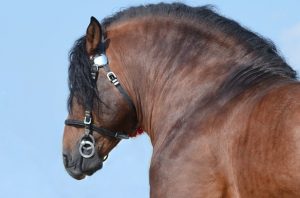
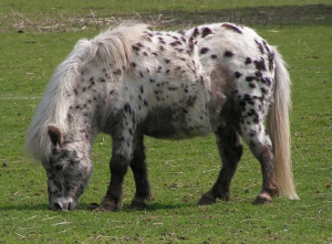
Most horse owners have heard about
equine Cushings disease and are familiar with some of the common clinical
signs: a long curly haircoat, delayed shedding, topline loss, pot-bellied
appearance, and sometimes laminitis. Additional signs include recurrent infections,
delayed healing, increased water intake and urination, and lethargy. Especially
as our equine companions live longer than ever before, a diagnosis of Cushings
disease is very common. But what causes these clinical signs and what can be
done for horses with this condition?
What is Equine Cushings/PPID?
Equine Cushings is the most common
endocrine disease in our older horse population. Cushings in humans and dogs
differs from the equine condition, so equine Cushings is more accurately known
as PPID (pituitary pars intermedia dysfunction).
The small pituitary gland is found
at the base of the brain near the hypothalamus. It is composed of three
different parts, each with unique functions. As indicated by the name, PPID
affects the pars intermedia portion of the pituitary gland. The neighboring
hypothalamus helps regulate the pituitary’s secretion of hormones. When this
regulation is disrupted, the pituitary continues to secrete hormones unchecked.
Usually the hypothalamus releases dopamine, a hormone, to signal the pars
intermedia to stop producing hormones. In older horses and PPID affected
horses, there is less dopamine to inhibit the pars intermedia, so it continues
releasing hormones and increases in size. The increased amounts of these
hormones, including one called ACTH, affect your horse’s thirst, thermoregulation,
and response to stress.
Diagnosis of PPID is made with
blood work to measure the level of the ACTH hormone. Completion of full
metabolic panel in addition to ACTH level is important to investigate other
hormones that may also be affected, such as insulin and leptin. One important
consideration is the time of year when blood is drawn, as ACTH levels of all
horses increase during the transitional fall period (approx. mid August through
end of November).
PPID Treatment Options
PPID is a manageable, but not
curable condition. The mainstay of treatment of PPID involves daily
administration of pergolide (prascend). This medication helps reduce ACTH
levels and improve clinical signs. Horse owners report improvement in shedding,
better maintenance/building of topline, and improved attitude/appetite. Treatment
of PPID can also help reduce circulating insulin levels in those horses that
experience elevated insulin secondary to PPID, thus reducing the risk of
laminitis. For horses with concurrent insulin dysregulation, additional
medications and dietary management may be indicated to further mitigate the
risk of laminitis. Many herbal remedies claim to aid in treatment of PPID, such
as chasteberry, but studies have failed to prove any benefits of supplementation.
Treating PPID with pergolide improves both quality and length of life for affected
horses.
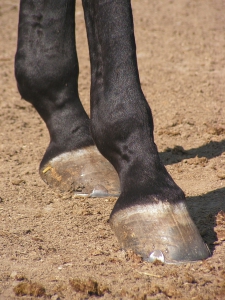
Thirty years ago, laminitis was usually a career-ending diagnosis, if not a life-ending one. Aside from mechanical support and pain management, little was known about the contributing factors associated with laminitis and the multitude of clinical factors that affect the severity, outcome, and likelihood of recurrence. Today, we know a great deal more about metabolic laminitis and specifically the role that insulin plays in mediating this disease process.
Current data shows that horses with an elevated baseline
insulin value are at a higher risk of developing hyperinsulinemic associated
laminitis (HAL). The ability to
establish a direct relationship between elevated serum insulin levels and the
onset of laminitis has enabled veterinary practitioners to make great strides
in combating this disease process.
Hyperinsulinemic Associated Laminitis Diagnosis and Treatment
When presented with a patient suffering from HAL, the first step is to determine just how high the insulin values are. Seasoned practitioners can usually estimate the severity of hyperinsulinemia by assessing the extent of regional adiposity (fat deposition in the horse’s body). The most common site of regional adipose tissue deposition are the crest of the neck, on either side of the withers, along either side of the horse’s topline, and on either side of the tail head. The greater the accumulation of fat in these areas, the higher the baseline insulin value in that patient. Bloodwork, in the form of a metabolic panel, is then used to quantify those observations to establish an appropriate therapeutic regimen and track progress throughout the treatment period.
If the patient is actively suffering from HAL, all
therapeutic methods are implemented in an attempt to slow down the damage
associated with the laminitic process.
In addition to dietary management and the eradication of starch from the
horse’s feed, medical intervention with metformin has proven to be a very
successful strategy in our practice.
Metformin increases tissue sensitivity to insulin in the
patient. Insulin is a signaling molecule which instructs cells to recover
glucose (starch/sugar) from the GI tract to use to power cellular
processes. In horses with
hyperinsulinemia, the tissues of the body aren’t responding to the insulin
currently being produced, so, the body produces more insulin, leading to a
hyperinsulinemic state.
The physiologic process by which elevated insulin values
lead to laminitis are still unknown.
However, current research shows that insulin is capable of binding to
receptors in lamellar epithelial cells which stimulates excessive growth of the
horn tubules, leading to the traditional elongated hoof structure of
chronically laminitic feet. Metformin
helps to increase tissue sensitivity to insulin which in turn down regulates
the body’s natural production of insulin.
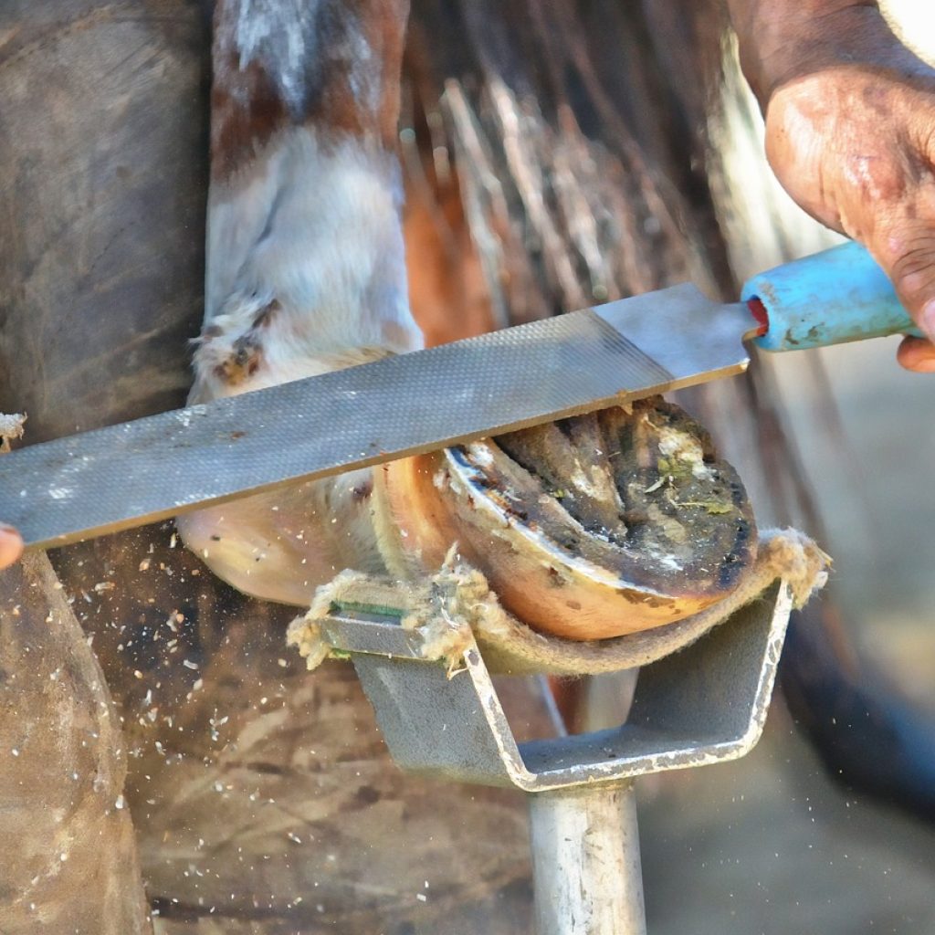
The success of treatment of horses with insulin dysregulation is highly dependent upon the severity of the HAL, the chronicity of the disease process, and the condition of the hoof capsule. Optimal outcomes are associated with high compliance on the part of the horse owner when it comes to implementing therapeutic regimens and dietary management, as well as the employment of a farrier who is willing to work with your veterinarian when making strategic decisions around trimming and shoeing your horse.
These patients require several series of radiographs over the course of their case in order to ensure optimal trimming is being performed in addition to tracking the sole depth of the patient. Most cases, when identified in their chronic stages, usually require many months to restore physiologic function of the hoof capsule and achieve an acceptable level of comfort on the part of the patient. Depending upon the integrity of the hoof capsule, metabolic stability, and comfort of the patient, these patients can sometimes return to their previous level of work. While not all cases have the perfect outcome, with the knowledge and medical advancements the veterinary profession has seen over the past few decades, it is absolutely worth trying to combat hyperinsulinemia associated laminitis.
Last night when you fed your horse you didn’t notice any
issues as you performed your quick “once over” before rushing back out the barn
door. But this morning they have a severely swollen leg, and they are hobbling
around.
What happened?
The list of possibilities isn’t terribly long, but there are
serious things on it: fractures, hoof abscesses, laminitis, joint infections,
and cellulitis. Of course, fracture is
the one that no one wants to hear and has significant ramifications. Laminitis and abscesses typically don’t have
the significant swelling described in the “case” above. Joint infections would likely have been
associated with trauma or a recent joint injection, and the swelling would
likely be associated with a particular joint, not the entire leg. That leaves this horse with a likely case of
cellulitis.
If you haven’t ever seen a case, they can be impressively swollen, and this can happen relatively quickly (overnight)! See below for an example.
What is cellulitis
Cellulitis is an infection of the deeper layers of the skin and the subcutaneous spaces. The pressure and inflammation make it extremely uncomfortable for the horse. They are usually very painful to the touch (maybe not everywhere they are swollen, but some portion of the swelling), and there is a lot of heat. Severe cases can have serum oozing from the skin. The horse may have a mild to moderate fever as well.
What causes cellulitis?
The infection can initiate from an obvious recent wound, bed sore, “scratches”, or other minor scrapes. It could also be a result of a more significant laceration from days before that seemed to be healing in the right direction. Another common route that these can begin is secondary to a hoof abscess. The infection begins in the hoof and then in the right set of circumstances, manages to take hold and cause further infection higher up the leg.
How is cellulitis treated?
If you notice the swelling before it is as extreme as the
picture above, getting medical attention could prevent it from becoming such an
advanced case. Medical attention usually
consists of antibiotics and anti-inflammatories systemically. On occasion, a “regional limb perfusion
(RLP)” may be justified. An RLP is a way
for the veterinarian to get very high concentrations of antibiotics to the area
of concern using a tourniquet and antibiotics directly infused into the
affected limb. Hydrotherapy (cold
hosing) is also often indicated.
Managing cases of cellulitis early is paramount to
preventing long term ramifications such as lymph damage (lymphangitis), or
laminitis. Lymphangitis is an inflammatory
process in the normal lymph drainage of the limb, and can leave the limb
permanently swollen. If there is enough
swelling in the leg during the cellulitic process, it could cause enough
vascular alterations to damage the laminae of the foot, causing laminitis.
If you find your horse with a sudden non-weight bearing
lameness (or barely weight bearing), we would always tell you that it is
justification for a phone call to your veterinarian, and most likely a visit.
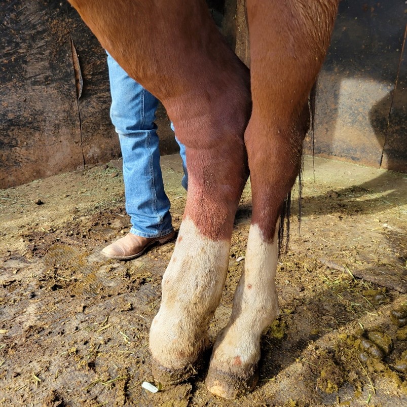
Sometimes when we as horseman see our horses on a routine basis, it can be easy to overlook that thickened, cresty neck or the fat pads that have developed on either side of their tail head. We recognize that it has been quite a while since we’ve been able to palpate any evidence of their ribs, but what other physical indicators do we use as veterinarians to assess the body condition of our patients? With the significant increase in metabolic laminitis cases seen on a national level, now more than ever, it is important to ensure our horses remain a healthy weight.
As a veterinarian, the first indication that I may be
dealing with a horse in metabolic crisis is not their enlarged abdominal girth
but instead is the thickened, hardened appearance to their crest. The crest is the region directly under the
horse’s mane along the topside of their neck.
A normal crest palpates soft, fluctuant, and homogenous to the
surrounding tissue and muscle. The crest
on a horse in metabolic crisis becomes hardened, like the density of
cauliflower. The crest can also become
wider and more visually pronounced in severe cases. As the metabolic disease process progresses,
horses can develop regional adiposity, also known as the accumulation of fat
pads throughout their body. These pads
of adipose tissue most commonly develop behind the shoulder blades on either
side of the withers and on either side of the tail head. In severe cases, adipose pads can develop along
either side of the thoracolumbar spine and in the girth region. A horse of a healthy weight has no evidence
of regional adiposity, with rib coverage that allows for palpation of each
individual rib but not the visual distinction of each rib.
Body condition scoring in horses is commonly done using the
Henneke System. This scoring system was
developed in 1983 and has been accepted throughout the international equine
industry as a standardized means to assess adiposity in horses. The scores range from 1 to 9 with a score of
1 being associated with extreme emaciation and a score of 9 correlating to
extreme obesity. Below is an outline of
the individual scores:
Poor: Grade 1 – Extreme
emaciation; no presence of fatty tissue, all bony prominences including
withers, shoulder blade, dorsal spinous processes, ribs, pelvis and sacrum are
all clearly visible.
Very Thin: Grade 2 –
Emaciated; slight tissue cover over bony prominences but withers, shoulder
blade, dorsal spinous processes, ribs, pelvis and sacrum are all clearly visible.
Thin: Grade 3 – Slight
accumulation of adipose tissue; bony prominences are no longer clearly
discernible; some fill over withers, shoulders and throughout the neck, ribs
still visible.
Moderately Thin: Grade 4 –
Ridge of spine and withers are still visibly pronounced, ribs still visible,
some accumulation of adipose tissue over the shoulders and through the neck and
tail head region.
Moderate: Grade 5 – Spine
and ribs cannot be visibly differentiated although ribs can be palpated, tail
head is soft and squishy; withers, shoulders and neck are smooth and rounded.
Moderately Fleshy: Grade 6 –
slight crease down the spine, tail head is still soft and squishy, regions of
adipose accumulation are present on either side of the withers and through the
crest, most ribs can still be palpated but not all of them.
Fleshy: Grade 7 – a crease
is present along the spine, the ribs have fat filling between them, tail head
is still soft, regions of adipose accumulation are pronounced through the crest
and on either side of the withers.
Fat: Grade 8 – a definite
crease is present along the spine (aka. “drainage ditch”), it is difficult to
feel any ribs, soft adipose tissue surrounds the tail head, the neck/crest is
quite enlarged and there is adipose accumulation on the inner aspects of the
hind limbs as well as behind the shoulder blades.
Extremely Fat: Grade 9 – the
“drainage ditch” collects water when it rains, there is bulging adipose
accumulation on top of the ribs, behind the shoulders, through the crest and on
either side of the tail head.
Accumulated abdominal fat is also present on the underside of the
flank.
To confirm your horse is in optimal health, it is important
to be able to accurately grade your horse’s body condition. An ideal range is between 4-6, depending on
the breed and usage of your horse. If
your horse falls outside of this range, it is important to discuss the
nutritional components of your horse’s diet with your veterinarian to decrease
the risks associated with equine obesity and ensure your horse isn’t deficient
in necessary nutrients.
At what age is a horse considered geriatric or senior? Does owning a senior horse mean that he must eat a feed labeled as a “senior” feed? As always, the answer is, it depends. Our horses are living longer lives than ever, thanks to an evolving understanding of the needs of geriatric horses and our ability to provide high quality care. There is no set age cutoff as to when a horse is considered geriatric, but most experts agree this can be around age 20. However, age is just a number, and many horses are still active and competing into their twenties, while others are happily retired. The main issues we see in our older horse population include dental disease, altered dietary needs, metabolic disease/PPID (Cushings), and arthritis.
Photo Courtesy PugnoM on Flickr
Dental Changes
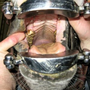
The ideal time to begin focusing on your horse’s regular preventative veterinary care is when he is young. Horses receiving regular preventative care, especially veterinary dentistry, preserve their teeth longer and can age more gracefully. Waiting until signs of dental disease or difficulty chewing are observed often indicates advanced disease and more limited treatment options. Most horses benefit from yearly dentistry, which includes a thorough sedated oral exam with a dental speculum, light source, and mirror. A thorough oral exam is vital to accurately assess, document, and treat any issues.
Horses suffering from abnormal dental wear, cracked or missing teeth, or severe periodontal disease may require more frequent care. Even though horse teeth are described as “long rooted,” they do eventually wear out, so proper care ensures the longest possible life of the tooth. Common dental issues in older horses include fracture of cheek teeth, complete expiration or wearing out of cheek teeth, loose/painful teeth, and periodontal disease. The incisors can also be affected by similar conditions.
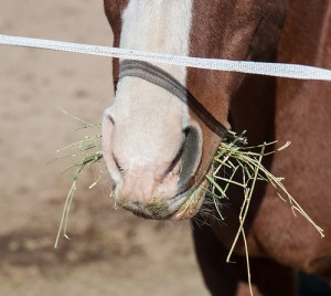
Staying on top of your senior horse’s dental care helps ensure that he gets what is needed from his diet. Sometimes severe dental disease and impaired chewing ability necessitates eliminating hay from the diet entirely. Complete feeds are formulated specifically for horses that can no longer chew hay effectively. These diets have greatly improved the quality and length of life for senior horses with compromised teeth.
Nutritional Changes
Nutrition recommendations for the geriatric horse are formulated around maintaining an ideal body condition. In the geriatric horse, this can be complicated by difficulty chewing, poor teeth, metabolic disease, and decreased ability to digest fiber and protein. For older horses in good weight and with adequate dentition, little dietary change may be required. For older horses who have difficulty maintaining weight and/or compromised teeth, complete feeds as mentioned above can help. These feeds are high in easily digestible fat, fiber, and protein, and are designed as easy-to-chew. They are formulated to replace hay entirely for those horses that can no longer adequately chew hay.
Adding water to soften the feed can reduce the risk of choke and ensure additional water intake. Feeding recommendations are often listed as pounds of feed per day depending on whether the horse is also eating hay, so it is important to weigh out your horse’s portion so that any necessary adjustments can be made more accurately.
Metabolic Changes
A curly haircoat and muscle loss over the back aren’t just signs of aging- They can indicate a common underlying condition: equine pars pituitary intermedia dysfunction, more commonly known as PPID or Cushings disease. This is a treatable (but not cureable) condition that is very common in geriatric horses, resulting in hormonal imbalances and symptoms such as delayed shedding, curly hair coat, muscle loss over topline, increased drinking and urinating, personality changes, and increased susceptibility to infections and laminitis. Diagnosis is made by a blood test as well as by history and clinical signs. Management involves daily administration of pergolide (Prascend) tablets, which help restore normal hormone levels and alleviate the clinical signs of the disease.
Equine Metabolic Syndrome (EMS) is a separate metabolic condition, but it sometimes occurs in conjunction with PPID. EMS horses typically show signs of being an “easy keeper”- cresty neck, regional fat deposits behind the shoulders and at the tail head. Horses with EMS are more susceptible to laminitis and usually have a higher baseline insulin level than normal. EMS horses are managed with strict attention to diet (minimizing sugar and starch) and exercise to help them maintain a lean body weight.
Joint and Muscle Changes
Just like us, aging horses suffer from daily aches and pains. An examination with your vet can help identify major and minor issues and
Photo Courtesy nikki_tate on Flickr
determine what treatment plan works best for you and your horse. Many options are available to keep our old horses comfortable.
Nutraceuticals (supplements) are very popular, although many of these products lack proof of efficacy and ingredients. More researched options include medications such as adequan (administered intramuscularly) and Legend (administered intravenously). These medications help provide the components necessary to keep joints healthy.
Joint injections target individual joints to reduce pain and inflammation and improve mobility. Non-steroidal anti-inflammatories such as Equioxx are also helpful, along with acupuncture and chiropractic care. Stable management is equally important- maximum turn out time allows horses to move at will, and monitoring herd dynamics ensures that older horses are not prevented from accessing food and water. In addition to these considerations, regular veterinary and farrier care will help your senior horse enjoy his golden years with you.
As always, if you have any questions about caring for your senior horse, please contact your veterinarian who can offer a personalized plan to help keep your geriatric horse happy, healthy, and comfortable well into their senior years.
As horsepeople, we know it’s necessary to implement slow transitions between grains and forages when changing our horse’s diet, but why the caution? Read on for the science behind slow feed changes.
Laminitis has been a bane of horseman and farriers since the horse was first domesticated. As our medical knowledge and diagnostic abilities have evolved, so has our understanding of the contributing factors of this disease process, including Equine Metabolic Syndrome. Equine Metabolic Syndrome is a term that’s been thrown around the equine community for the past few decades, but what does it mean?
What is EMS?
Equine metabolic syndrome (EMS) is a condition most commonly characterized by an inability to properly metabolize carbohydrates. The disease has been known by many names, including hypothyroidism, peripheral Cushing disease and pre-laminitic syndrome. Today, we know that Equine Metabolic Syndrome is characterized in horses, ponies and donkeys by obesity, regional deposition of fat, and systemic insulin resistance.
Insulin is an important hormone that allows the cells to obtain glucose from food. Without insulin, or a proper response to insulin, cells cannot utilize glucose, regardless of how much food the horse is fed. This starvation process at the cellular level pushes the horse into a stressed state (characterized by elevated cortisol levels) where the body is encouraged to hold on to as much fat as possible, making weight loss next to impossible in these horses. In addition, glucose deprivation in the laminae of the horse’s feet can lead to laminitis. How is it Diagnosed?
When we suspect a horse may have EMS, we will recommend bloodwork to confirm our suspicions as well as guide us in the proper treatment of your horse. Horses with EMS may also have concurrent Cushing disease, which has been shown to be a contributing factor to the initial development of EMS. It is important for us to understand the underlying metabolic factors that are contributing to your horse’s case in order to provide the most efficacious recommendations both medically and diet.
The key values we assess when running blood work for EMS include:
ACTH: When elevated, this value can be indicative of Equine Cushing’s Disease.
Insulin: Elevated levels of insulin indicate insulin resistance.
Leptin: This is a secondary value, used to ascertain the validity of elevated Insulin. When leptin is elevated in addition to elevated insulin, EMS and IR (insulin resistance) are valid diagnoses.
Glucose: When both ACTH and insulin are elevated, glucose can help differentiate the primary disease process.
What Can You Do About It?
Horses that are merely overweight but not actively laminitic should be put on a low starch diet, comprised of hay with less than 12% non-structural carbohydrates fed at 1-1.5% body weight. (in this example, a 1000lb horse should be fed 10-15lbs of hay per day). Their forage diet can be adequately balanced using either a ration balancer or a specifically formulated low starch feed. These horses also benefit from routine exercise as an additional aid for weight loss.
In those patients that are actively laminitic, exercise is not recommended but the same dietary recommendations apply. In addition, if your horse is diagnosed as insulin resistant (IR), metformin is a beneficial medication used to increase tissue sensitivity to insulin. Horses with IR are comparable to humans with type 2 diabetes, in that they produce adequate amounts of insulin, but their body simply fails to respond appropriately. Metformin is a medication commonly used in humans with IR and its efficacy in equine IR cases has been confirmed both in laboratory and field studies.
Another option to stimulate weight loss in EMS horses is levothyroxine, commonly sold in the equine community as Thyro-L. Thyro-L functions by increasing the rate of the horse’s metabolism to further stimulate weight loss. It is important to understand that Thyro-L has no direct impact on the laminae of an actively laminitic horse and instead works indirectly over a longer period of time.
EMS is a frustrating disease for practitioners, clients and patients alike. The more we understand of the disease process the better our chances at restoring metabolic equilibrium and preventing further deterioration of the laminae. If you suspect your horse may have EMS, talk to your vet about appropriate diagnostic measures.
Laminitis. Founder. Words no horse owner wants to hear. But is it a death sentence for your horse? Or can they come back from it? Find out more in this month’s blog post.